
Treating Supracondylar Elbow Fractures in Children: Seeing the Challenges, Heading Off the Risks
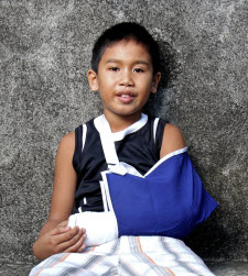 For young children, running, climbing, and jumping are a normal part of healthy development. With such activities, however, comes potential for injury. According to Douglas Armstrong, MD, Penn State Bone and Joint Institute, “Pediatric supracondylar fracture is common in kids 4 to 8 years old when they fall from monkey bars or other climbing equipment.”
Armstrong cautions against trying to reduce such fractures in the emergency department (ED) with the patient awake. “Severely displaced fractures present with pain, swelling and obvious deformity. The diagnosis is made with physical exam and plain radiograms. Most displaced fractures are treated with closed reduction in the operating room under general anesthetic with complete relaxation. Fixation with two or three K-wires allows the elbow to be immobilized in moderate flexion, reducing the risk of vascular embarrassment.”
The majority of children recover without problem. Nevertheless, Armstrong notes, “When elbow fracture complications do occur, they can be serious. Nerve injuries may be seen in 25 percent of extension type supracondylar fractures and 30 percent of flexion types. In children, these may be impossible to diagnose acutely. Every effort should be made to perform a neurovascular examination. Most such injuries are neurapraxia and resolve on their own over the course of weeks.
Occasionally nerve dysfunction can signify entrapment of the nerve or artery. Inability to obtain fracture reduction in the context of a nerve injury can indicate need for open reduction. Acute vascular compromise, although uncommon, requires emergent treatment. Flow through the brachial artery can be blocked as a consequence of entrapment, intimal tear, or arterial rupture. The signs of loss of perfusion to the hand are well known. Treatment in the ED may include gently flexing the elbow about 10 to 15 degrees to relieve tension on the anterior neurovascular structures. Emergent reduction in the operating room (OR) with fixation of the fracture is required. There is controversy regarding the optimal treatment for a hand that remains pulseless after anatomic reduction. At Milton S. Hershey Medical Center, when the hand is pink we generally treat with expectant monitoring, but arterial exploration and reconstruction is an option.”
Acute compartment syndrome (CS) is rare, but is one of the most feared complications of supracondylar fracture. The usual signs and symptoms of CS in a young child can be subtle or even absent and the condition can develop insidiously, with devastating consequences. Especially in a young child with a badly displaced supracondylar fracture, surgeons and nurses need to be alerted to increased requirement for analgesia, agitation, progressive loss of sensory and or motor function.
Armstrong advises, “Less severe supracondylar fractures can present with a painful elbow with little swelling and no deformity. Careful physical exam can localize the site of injury. When in doubt, comparison X-rays can be helpful. If the distal fragment tilts, cubitus varus can develop. The orthopaedic surgeon needs to be alert to clinical and radiographic signs of rotation or tilting of the distal fracture fragment. Careful clinical exam of the carrying angle and high quality orthogonal radiograms are needed. If cubitus varus develops, the outcomes of contemporary corrective osteotomy are satisfactory for most.”
After fixation of the fracture, the child often goes home within twenty-four hours. Pins are usually removed at three weeks in the clinic; anesthesia is not required. Generally most children recover without physical therapy and return to their usual activities at nine weeks after injury.

Douglas G. Armstrong, MD
Edwards P. Schwentker Professor
Orthopaedics Director, pediatric orthopaedic surgery
Phone: 717-531-4826
Email: darmstrong@pennstatehealth.psu.edu
Fellowship: Pediatric orthopaedics, Buffalo Children’s Hospital, Buffalo, NY; Spine surgery, University of Ottawa Civic Hospital, Ottawa, Ontario
Residency: Orthopaedic surgery, McGill University, Faculty of Medicine, Montreal, Quebec
Medical School: University of Ottawa Faculty of Medicine, Ottawa, Ontario
Connect with Douglas G. Armstrong, MD, on Doximity
U.S. News & World Report 2025-2026
Penn State Bone and Joint Institute represents the clinical aspect of Penn State Orthopaedics and Rehabilitation. Faculty from Penn State Orthopaedics and Rehabilitation teach and perform leading-edge research in the department, and provide innovative care in the multi-disciplinary Penn State Bone and Joint Institute. As a department, Penn State Orthopaedics and Rehabilitation faculty, clinicians and researchers have been recognized nationally, ranking twentieth in orthopaedic research funding from the National Institutes of Health.
Continuing Education activities
Stay informed of all Continuing Education activities that interest you.